EYE ANATOMY
Don’t remember the lessons on eye anatomy from your high school biology class? That’s OK—we have provided the following eyeball illustration and terms just to give you a refresher course. And we won’t give you a pop quiz afterwards…
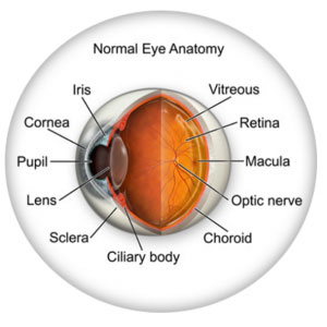
CORNEA: |
Transparent front segment of the eye that covers iris, pupil, and anterior chamber, and provides most of an eye’s optical power. |
PUPIL: |
Variable-sized, circular opening in center of iris; it appears as a black circle and it regulates the amount of light that enters the eye. |
IRIS: |
Pigmented tissue lying behind cornea that (1) gives color to the eye, and (2) controls amount of light entering the eye by varying size of black pupillary opening; separates the anterior chamber from the posterior chamber. |
LENS: |
Natural lens of eye; transparent intraocular tissue that helps bring rays of light to focus on the retina. |
RETINA: |
Part of the eye that converts what we see into electrical impulses sent along the optic nerve for transmission back to the brain. Consists ofmany named layers that include rods and cones. |
MACULA: |
Small, specialized central area of the retina responsible for the sharpest central vision. |
VITREOUS: |
Transparent, colorless, gelatinous filling; in the rear two-thirds of the interior of the eyeball, between the lens and the retina. |
OPTIC NERVE: |
Largest sensory nerve of the eye; carries impulses for sight from retina to brain. |
SCLERA: |
The white of the eye; a protective fibrous layer that is the outer covering of the eyeball except for the part that is the cornea. |
CILIARY BODY: |
A muscular ring under the surface of the eyeball; helps the eye focus by changing the len’s shape and also produces aqueous humor. |
CHOROID: |
The vascular layer between the sclera and the retina; the blood vessels in the choroid help provide oxygen and nutrients to the eye. |
VISION CONDITIONS
AMBLYOPIA
Amblyopia, commonly called lazy eye, occurs when one eye develops differently than the other eye does, causing one eye to be weaker than the other. Sometimes a difference in focusing ability causes one eye to be used more often. Other times, the eyes are misaligned, causing one eye to “shut off” to avoid double vision. Regardless of the cause, the result is a weakened, or amblyopic, eye.
SYMPTOMS
It’s hard to spot amblyopia. Sometimes a child will noticeably favor one eye over the other. Another possible symptom is the child frequently bumping into things on one side. The best way to tell if your child has lazy eye is through a complete exam at about six months and three years. Early diagnosis can prevent amblyopia from leading to more serious problems, such as loss of the ability to see three dimensions or functional blindness in the amblyopic eye
ASTIGMATISM
Sometimes the cornea is irregularly shaped, causing the eye to focus an object on two different areas of the retina. This is known as astigmatism. For the cornea to bend light correctly, it should be dome-shaped, like a basketball. Astigmatic corneas are shaped more like a football. This causes a distorted view when looking at objects which are close-up and far away.
The cause of astigmatism is unknown, but we should remember that astigmatism is NOT an eye disease, but an eye condition which is fairly common. Astigmatism is often associated with myopia or hyperopia, and it usually is present from birth. It may be hereditary, or it may be caused by factors such as pressure on the cornea, incorrect posture, or increased use of the eyes for “near work.”
Mild astigmatism may not need to be corrected. Eyeglasses, contact lenses, or refractive surgery can correct moderate to high degrees of astigmatism.

COMPUTER VISION SYNDROME
Computer vision syndrome (CVS) affects three out of four computer users. It is a series of symptoms related to extended periods of computer usage. Though it is no cause for panic, measures can be taken to relieve symptoms of CVS.
SYMPTOMS
CVS can appear as a variety of symptoms. Headaches, eye strain, neck and back aches, sensitivity to light, blurred vision, double vision, and dry or irritated eyes are all possible problems related to CVS.
EMMETROPIA
When rays are focused correctly on the retina of a relaxed eye, the eye is said to be emmetropic. Emmetropia is the medical term for 20/20 vision, vision that needs no corrective lenses, contact lenses, or reading glasses. It occurs because the optical power of the eye can perfectly focus an image to the retina, giving it “perfect” vision.
The opposite of emmetropia is ametropia. With ametropia, the focal point of the eye is some distance in front of or behind the retina. The following vision conditions are types of ametropia.
HYPEROPIA (FARSIGHTEDNESS)
Hyperopia is more commonly known as farsightedness. As the name suggests, people with farsightedness are able to focus on objects that are further away, but have difficulty focusing on objects which are very close. This is because the eyeball is shorter than normal, which prevents the crystalline lens in the eye from focusing correctly on the retina. About a fourth of the population is farsighted. Hyperopia can lead to chronic glaucoma, a more serious condition, later in life.
A family history of hyperopia is a risk factor for developing hyperopia. Babies are often born with hyperopia but they can usually outgrow the condition as their eyes develop into the correct shape.
Hyperopia can be corrected with eyeglasses or contact lenses. There are also new surgical procedures that can correct hyperopia.

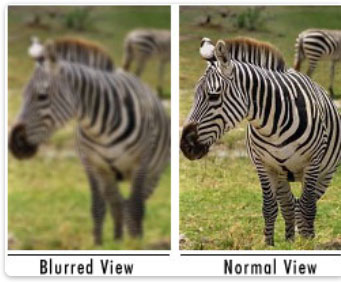
MYOPIA (NEARSIGHTEDNESS)
Myopia is the medical term for what most people call nearsightedness. It is a condition where you can see objects clearly only when they are closer, but when objects are further away you can’t focus on them. Myopia usually develops in early childhood, though it sometimes develops in early adulthood. In rare cases, myopia can lead to more serious conditions such as retinal detachment.
Myopia is considered a genetic disorder. If your parents are nearsighted, you are at greater risk of also being nearsighted. Another risk factor is ‘near work’ – work involving fine detail or focusing on close objects.
Myopia can be accommodated and sometimes corrected with eyeglasses or contact lenses. Sometimes myopia continues to gradually worsen throughout life, a condition known as myopic creep. Myopia can also be corrected by LASIK surgery.

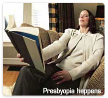
PRESBYOPIA
As a people get older, usually when they hit their mid to late 40s, a condition called presbyopia can set in. Presbyopia is the inability to focus on objects near the eye. Mother nature essentially shuts our auto focusing mechanism down. One usually notices that it is harder to read or use the computer. Bifocals or reading glasses are a way to remedy this condition.
Presbyopia is a natural consequence of the aging process. There is no cure, though researchers are constantly looking for one. Even if someone has never had vision problems before, he can still develop presbyopia. It may seem to occur suddenly, but it actually occurs over a long period of time. Symptoms include having to hold things at arm’s length to see them clearly, eye strain, fatigue, and headaches from near work.
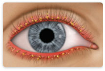
BLEPHARITIS
There are two types of blepharitis. Seborrheic blepharitis is often part of an overall skin condition called seborrhea, which may also affect the scalp, chest, back and the area behind the ears.
The second form of blepharitis – staph blepharitis – is a more common condition, caused by bacteria, that begins in childhood and may continue through adulthood.
SYMPTOMS
Blepharitis could be described as dandruff of the eyelids. Seborrheic blepharitis causes redness of the eyelids, flaking and scaling of the eyelashes, and greasy, waxy scales. Staph blepharitis also causes redness of the eyelid margins and flaking of the lashes, and can cause loss of eyelashes, eyelid scarring, and red eye.
CATARACTS
A cataract is a cloudiness that occurs in the lens inside of the eye. A cataract is a cloudiness that occurs in the lens of the eye. The lens is made mostly of water and protein that is arranged to let light through. Sometimes the protein clumps, blocking light and making the lens appear cloudy.
SYMPTOMS
A person with cataracts may encounter faded colors, problems with light (such as halos, or headlights that seem too bright), poor night vision, double vision, or multiple vision.
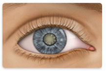
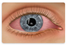
CONJUNCTIVITIS (PINK EYE)
Conjunctivitis, commonly called pink eye, is a redness of the eye. It is often accompanied by a discharge (clear, yellow, or white) and itching in the eye. Pink eye is most often a viral infection, but it may also be caused by bacteria or an allergic reaction. Viral pink eye is highly contagious.
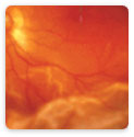
DIABETIC RETINOPATHY
Diabetic retinopathy is a condition associated with diabetes. High levels of blood sugar may damage tiny blood vessels in your eye. New vessels may form to replace the damaged vessels. The new vessels can burst, resulting in blurred vision or even blindness.
SYMPTOMS
Symptoms of diabetic retinopathy include:
“Floaters” – small specks that pass across your field of vision, made up of cells floating in the transparent gel of your eyeball Difficulty reading or seeing things close-up Sudden loss of vision Flashes Blurred or darkened vision
DRY EYE SYNDROME
If your eyes are constantly itchy or dry, you may have dry eye syndrome, which affects almost 10 million Americans. Dry eye syndrome is caused by a lack of, or poor quality of, tears. Tears lubricate the outer layer of the eye called the cornea. If the tears are not composed of a proper balance of mucous, water, and oil, the eye becomes irritated.
SYMPTOMS
Dry eye syndrome leads to a number of symptoms, including itching, irritation, burning, excessive tearing, redness, blurred vision that improves with blinking, and discomfort after long periods of watching television, driving, using a computer, or reading.
GLAUCOMA
Glaucoma is a very common eye disorder affecting millions of Americans. It is caused by too much pressure on the inside of the eye. The fluid in your eyes helps to nourish and cleanse the inside of your eyes by constantly flowing in and out. When the fluid is prevented from flowing out, the intraocular pressure builds and damages the optic nerve. This causes a gradual loss in peripheral vision.
SYMPTOMS
Those suffering from open-angle glaucoma experience a type of tunnel vision, where their field of vision gradually decreases. It can eventually lead to blindness. Narrow-angle glaucoma, which is rare, carries symptoms of sharp pain in the eyes, blurred vision, dilated pupils, and even nausea or vomiting. It can cause blindness in a matter of days, and it requires immediate medical attention.
MACULAR DEGENERATION
Macular degeneration is a disease which affects a small area of the retina known as the macula. The macula is a specialized spot on the retina that allows us to see the fine detail of whatever is directly in front of us. Macular degeneration occurs when the macula begins to deteriorate. Most often, macular degeneration is accompanied by formation of yellow deposits, called “drusen,” under the macula, which dry out or thin the macula. This is called “dry” macular degeneration. In rare cases, abnormal blood vessels develop under the macula and leak fluid. This is called “wet” macular degeneration.
SYMPTOMS
It is difficult to detect dry macular degeneration in its early stages. The most common symptoms, when detected, include a spot of blurry vision, dark vision, or distorted vision. Wet macular degeneration progresses much faster than the dry variety. Both forms of macular degeneration can cause blindness.
RETINAL DETACHMENT
The part of the eye which collects light and transmits the images to the optic nerve and brain is the retina. It lines the inner back wall of the eye. When separated from the back wall, it is known as retinal detachment.
SYMPTOMS
A retinal detachment may cause a sudden defect in your vision. It may just cause a blind spot too small to notice, or it may cause a noticeable shadow which obscures your vision. A sudden increase in “floaters,” which look like small particles or fine threads, may also be noticed. Finally, flashes of light may be associated with retinal detachment.